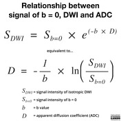
mri b value
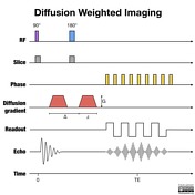
Part of the MR Diffusion Functional Group Macro with usage. The b value is used in MRI in the context of Diffusion Weighted Imaging DWI.
Diffusion Weighted Imaging Radiology Reference Article Radiopaedia Org
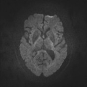
A b factor of zero b 0 smm 2 indicates no diffusion weighting and the image is analogous to a T2-weighted image.

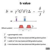
. B value measures the degree of diffusion weighting applied thereby indicating the amplitude G time of applied gradients δ and duration between the paired gradients Δ and is calculated as. B γ² G² δ² Δδ3 Therefore a larger b value is achieved by increasing the gradient amplitude and duration and by widening the interval between paired. Depending on the organ being imaged b-values typically range from 50-1000smm 2.
Constant b in GutenbergRichter law. DWI is done to determine the rate of molecular diffusion in different areas of the body. 2015 designed a study to correlate the accuracy of 3T MRI in which DWI occurred with a b-value of 2000smm 2.
The value of b was set at 0 30 50 80 100 150 200 400 600 800 smm 2 and varied throughout a single imaging session. Our purpose was to determine whether high-b-value diffusion-weighted MR imaging improved contrast and detection of signal changes in acute and chronic. The best b-value combination was 0 and 600 smm2 and multiple b2.
In the abdomen lower b values applied often range from 50 to 150 smm 2. Studies have reported that the use of b values higher than 1000 smm 2 and 2000 smm 2 improves tumor localization and the contrast between benign and malignant lesions in the prostate and the breasts 12 14. Two recent studies explored DWI at an ultra high b-value of 2000smm 2.
Reported that pre-treatment ADC and a diffusion index RD can both correlation well r 076077 with brain tumor response to radiation therapy 87. With enhanced gradients the whole b ra in can b e scanned with in seconds. 335 ms producing b-values of 1018 2475 3069 4009 5911 818111295 15750 smm2.
The strong correlation implies that tumors with low. Recent technological advances in MR instrumentation allow acquisition of whole-brain diffusion-weighted MR scans to be obtained with b values greater than 1000. If the relationship between MR signals and b-values were monoexponential the ADC values would remain constant for any 2.
In general approximately 1000 smm 2 is the maximal b value for DWI 5 11. Beside the diffusion-weighted row data images there is one more way to present diffusion data which is called the apparent diffusion coefficient ADC map of water. In general in healthy tissue molecules of water and other chemicals are not stationary but moving about.
Using a Gaussian model with b-values up to 4000 smm 2 Mardor et al. Multiple b values of 600 smm2 and higher are recommended to differentiate between benign and malignant. These can either be calculated directly from the isotropic DWI images or by finding the arithmetic mean of ADC values generated from each directional diffusion map.
Conventional MRI and multi- b -value DW images were collected before treatment at mid-stage evaluation and when conducting therapeutic efficiency assessment after chemotherapy. Acquisition parameter in diffusion MRI. B-value may refer to.
One hundred six patients underwent diagnostic multiparametric prostate MRI at 3T using an endorectal coil. 5 TR 2500 ms TE678ms and NEX4 averaged off line. High b-values with or without non-Gaussian models have been used for early evaluation of cancer treatment.
The degree of diffusion weight in g correlates with the strength of the diffusion gradients characterized b y the b - value which is a function of the gradient related parameters. ADC values decrease when b-values are increased beyond 1000 smm 2. The higher the b values the better the sensitivity of diffusion weighted imaging usually three to four b values are used in diffussion-weighted sequences b50 b500 b1000 and b1400.
In DWI we recommend the use of b-values of 0 and 800 smm2 as two b-values or b0 50 600 800 and 1000 smm2 as multiple b-values for distinguishing between benign and malignant liver lesions. BACKGROUND AND PURPOSE. 5 b-value b 0 188 375 563 750 smm2 DWI and high b-value b0 1000 and 2000 smm2 DWI were acquired.
Strength duration and the period b etween diffusion gradients. May be present otherwise. Calculated high b-value b1000 smm2and b2000 smm2 DWI were derived from the DWI dataset using DK and IVIM models.
In contrast this molecular motion may be obstructed in certain pathological conditions - such as in the. 27 ms big delta. Important characteristic of a thermistor B parameter B-factor in crystallography.
The lesionnormal parenchymal ADC ratio for b600 b1000 and multiple b2 better distinguished between benign and malignant lesions. Required if Frame Type 00089007 Value 1 of this frame is ORIGINAL. Diffusion weighted gradient strength Gmax has been increased in steps of 29 44 49 56 68 80 111 mTm small delta.
Thirty-five PCa patients who were to be treated with radical prostatectomy underwent 3T DWI-MRI. Higher b values typically range between 500 and 1000 smm 2. The ADC map in contrast is related to the natural logarithm ln of the isotropic DWI divided by the initial T2 signal b0.
Diffusion-weighted MRI and optimal b-value for characterization of liver lesions. 16 2731 DeLano et al 16 reported that ADC values decreased by approximately 30 to 35 when b-values were increased from 1000 smm 2 to 3000 smm 2 for some ROIs. Topics referred to by the same term.
This is the actual b-value for original frames and those derived from frames with the same b-value or the most representative b-value when derived from images with different b-values. In DWI the optimal b value is 600 smm2.
2
Apparent Diffusion Coefficient Radiology Reference Article Radiopaedia Org

Diffusion Weighted Imaging Radiology Reference Article Radiopaedia Org

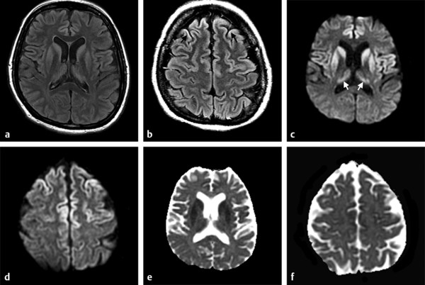
Conventional Mri Dwi And Adc Images Of Braf V600e Mutant And Download Scientific Diagram

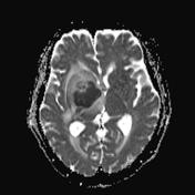
A Mri With A Normal Brain B Mri With A Brain Tumor 13 Download Scientific Diagram
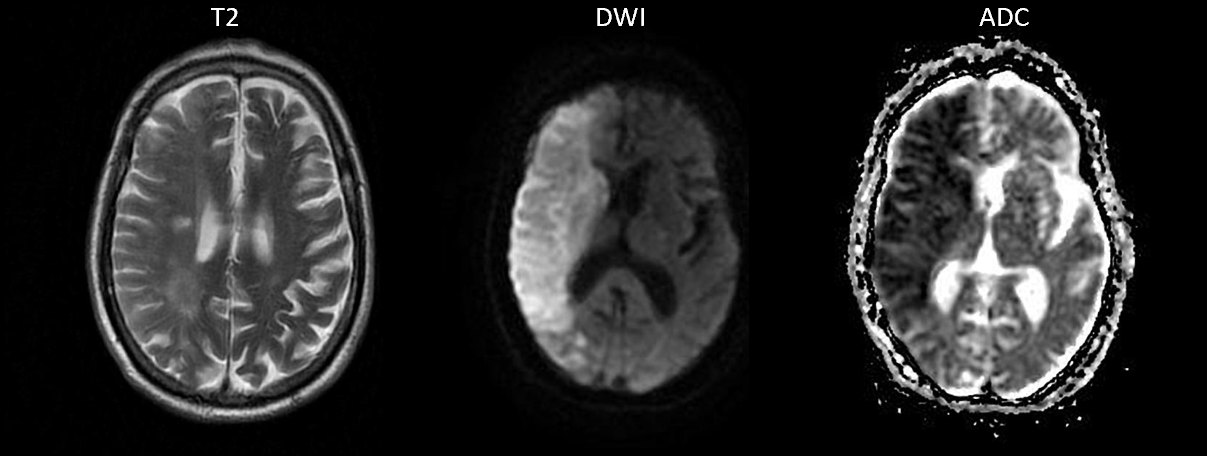
Signal Intensity Of Dwi And Adc In Diffusion Restriction Increased Download Scientific Diagram
Diffusion Weighted Imaging In Hemorrhage Radiology Key

Mri Planes For Mri Head Scan A Axial B Coronal C Sagittal Mr Download Scientific Diagram
Magnetic Resonance Imaging Mri And Computerized Tomography Download Scientific Diagram
Tensor Valued Diffusion Encoding For Diffusional Variance Decomposition Divide Technical Feasibility In Clinical Mri Systems Plos One

Apparent Diffusion Coefficient Radiology Reference Article Radiopaedia Org
The Basics Of Mri Interpretation Radiology Geeky Medics
2
Diffusion Weighted Imaging Radiology Reference Article Radiopaedia Org

Principles Of Diffusion Tensor Imaging And Its Applications To Basic Neuroscience Research Neuron

Apparent Diffusion Coefficient Radiology Reference Article Radiopaedia Org
Diffusion Weighted Imaging Radiology Reference Article Radiopaedia Org
Diffusion Weighted Imaging Radiology Reference Article Radiopaedia Org

Apparent Diffusion Coefficient Radiology Reference Article Radiopaedia Org


Comments
Post a Comment